Female abnormal lumbar MRI scans are a crucial tool in diagnosing and understanding lower back conditions in women. Lumbar MRI (Magnetic Resonance Imaging) provides detailed images of the spine, intervertebral discs, nerves, and surrounding tissues, allowing healthcare professionals to identify structural abnormalities, degenerative changes, or injuries. Women may present with a variety of lumbar spine issues, ranging from herniated discs to spinal stenosis, spondylolisthesis, or congenital anomalies. Early detection of abnormalities through lumbar MRI can guide treatment decisions, reduce the risk of complications, and improve overall quality of life.
Understanding Female Lumbar Spine Anatomy
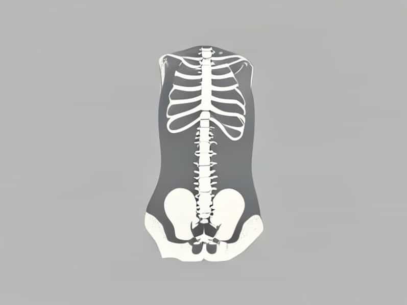
The female lumbar spine consists of five vertebrae (L1-L5), intervertebral discs, ligaments, muscles, and nerves that support body weight, enable movement, and protect the spinal cord. The unique anatomy of women, including differences in pelvis structure and ligamentous laxity, may influence susceptibility to certain lumbar conditions. Hormonal factors, particularly estrogen levels, can also affect bone density, disc health, and joint stability, making lumbar MRI an important diagnostic tool in evaluating spine health in women.
Key Anatomical Features
- Lumbar vertebrae (L1-L5) providing structural support and flexibility.
- Intervertebral discs acting as cushions and shock absorbers.
- Spinal nerves transmitting signals between the spine and lower body.
- Ligaments and muscles stabilizing the spine and maintaining posture.
- Pelvic alignment influencing lumbar curvature and load distribution.
Common Abnormalities in Female Lumbar MRI
Female patients undergoing lumbar MRI may present with a range of abnormalities. These can include degenerative changes, disc herniation, spinal stenosis, spondylolisthesis, and congenital or acquired deformities. Abnormalities are often linked to symptoms such as chronic lower back pain, leg pain, numbness, tingling, or weakness. Identifying these changes accurately through MRI is critical for planning effective interventions and preventing long-term disability.
Degenerative Disc Disease
Degenerative disc disease is a common abnormality seen in female lumbar MRI, often related to aging, repetitive stress, or hormonal changes. The intervertebral discs lose hydration and elasticity, leading to reduced disc height and increased risk of herniation or nerve compression. Women may experience chronic lower back pain, stiffness, or radiating leg pain as a result of these changes. MRI can reveal disc dehydration, bulging, or fissures, which are essential for diagnosis and treatment planning.
Herniated Discs
Herniated discs occur when the inner gel-like nucleus of a disc protrudes through the outer fibrous ring, potentially compressing nearby nerves. Women may develop lumbar disc herniation due to lifting injuries, prolonged sitting, pregnancy-related spinal stress, or degenerative changes. Lumbar MRI can identify the location, size, and severity of herniation, which helps guide conservative management or surgical interventions when necessary.
Spinal Stenosis
Spinal stenosis involves narrowing of the spinal canal or foramina, leading to nerve compression. Female patients may experience symptoms such as lower back pain, leg pain, or difficulty walking. Lumbar MRI provides a clear view of canal dimensions, ligament hypertrophy, or disc protrusions that contribute to stenosis. Identifying stenosis early is crucial to prevent nerve damage and improve mobility through targeted treatments.
Spondylolisthesis
Spondylolisthesis is the forward slippage of one vertebra over another, which can occur due to congenital factors, degenerative changes, or trauma. Women with spondylolisthesis may report chronic lower back pain, radiating leg pain, or postural changes. MRI is effective in visualizing vertebral alignment, disc integrity, and associated nerve compression, aiding in decisions regarding physical therapy, bracing, or surgery.
Other Potential Findings
Aside from degenerative or structural changes, female lumbar MRI can reveal other abnormalities, such as vertebral fractures, tumors, infections, or inflammatory conditions. These findings require careful evaluation and correlation with clinical symptoms to determine appropriate management. For example, osteoporosis-related vertebral fractures are more prevalent in postmenopausal women, highlighting the importance of MRI in detecting subtle fractures that may not be apparent on X-rays.
Additional Findings on MRI
- Osteoporotic vertebral compression fractures.
- Spinal tumors or cysts.
- Inflammatory or infectious conditions like spondylitis or discitis.
- Congenital anomalies such as spina bifida occulta.
- Post-surgical changes in patients with previous spine operations.
Clinical Symptoms Associated with Abnormal MRI Findings
Female patients with abnormal lumbar MRI often present with a variety of symptoms. Common complaints include chronic low back pain, sciatica, numbness, tingling, and weakness in the lower extremities. Symptoms may worsen with activity, prolonged standing, or sitting and can interfere with daily life. MRI findings allow physicians to correlate symptoms with structural abnormalities, guiding appropriate interventions such as physical therapy, medication, or surgery.
Typical Symptom Patterns
- Localized lower back pain exacerbated by movement.
- Radiating leg pain, commonly known as sciatica.
- Numbness or tingling in the legs or feet.
- Muscle weakness affecting mobility or balance.
- Postural changes or reduced range of motion due to pain.
Importance of Early Diagnosis and Intervention
Early detection of abnormalities on lumbar MRI is critical in preventing long-term complications and improving quality of life for female patients. Prompt diagnosis allows for tailored treatment strategies that may include lifestyle modifications, targeted physical therapy, pain management, or surgical interventions. Regular monitoring and follow-up imaging may be recommended for progressive conditions, ensuring that treatment remains effective and complications are minimized.
Benefits of Early Intervention
- Prevention of chronic pain and disability.
- Improved mobility and functional outcomes.
- Reduced risk of nerve damage or paralysis in severe cases.
- Opportunity for non-invasive treatments before surgery becomes necessary.
- Enhanced overall quality of life through timely management.
Female abnormal lumbar MRI is an essential diagnostic tool that provides detailed insights into spinal health, structural abnormalities, and degenerative changes. Recognizing the significance of MRI findings allows healthcare providers to develop targeted treatment plans, prevent complications, and improve patient outcomes. Women may experience a range of lumbar spine conditions influenced by anatomy, hormonal factors, and lifestyle, making individualized assessment and management critical. Early detection, careful evaluation, and appropriate intervention are key to maintaining spinal health and ensuring long-term well-being.