The parathyroid glands are small yet vital components of the endocrine system, playing a crucial role in regulating calcium levels and overall metabolic balance within the human body. Despite their small size, these glands have a complex structure and sophisticated functionality that significantly impact bone health, kidney function, and neuromuscular activities. Understanding the structure of the parathyroid gland provides insight into how it performs its essential functions, how it interacts with other endocrine organs, and why it is critical in maintaining calcium homeostasis. Knowledge of its anatomy and cellular composition is also fundamental for medical professionals diagnosing and treating disorders such as hyperparathyroidism and hypoparathyroidism.
Anatomical Location of the Parathyroid Glands
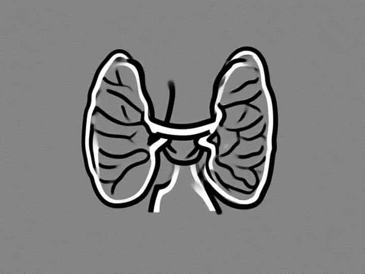
The parathyroid glands are typically four small, oval-shaped glands located on the posterior surface of the thyroid gland in the neck. They are usually paired, with two glands on each side, referred to as the superior and inferior parathyroid glands. Their exact location can vary among individuals, with the superior glands generally situated near the upper poles of the thyroid lobes and the inferior glands near the lower poles. Despite their small size, usually measuring about 6 millimeters in length and 3-4 millimeters in width, their strategic location allows them to efficiently monitor and regulate blood calcium levels in close proximity to the thyroid and major vascular structures.
Blood Supply and Innervation
The parathyroid glands have a rich blood supply primarily derived from the inferior thyroid arteries, with some contribution from the superior thyroid arteries. This extensive vascular network is essential for the rapid release of parathyroid hormone (PTH) into the bloodstream. Venous drainage occurs through small veins that eventually drain into the thyroid venous plexus. Innervation of the parathyroid glands comes from the cervical sympathetic ganglia, which helps modulate blood flow but does not directly control hormone secretion. The combination of rich vascularization and sympathetic innervation ensures that the glands respond promptly to changes in serum calcium levels.
Microscopic Structure of the Parathyroid Gland
Microscopically, the parathyroid gland consists of densely packed cells organized into cords, clusters, and follicles. The main functional units are the parenchymal cells, which are primarily responsible for hormone production. These cells are surrounded by a delicate connective tissue framework containing blood vessels, lymphatics, and nerves. The structure is optimized to allow rapid sensing of calcium levels and prompt hormone secretion.
Cell Types
There are two main types of cells within the parathyroid gland, each with distinct roles in endocrine function
-
Chief CellsChief cells are the most abundant and are primarily responsible for the production and secretion of parathyroid hormone (PTH). These cells have a granular cytoplasm rich in secretory vesicles and are highly sensitive to changes in serum calcium levels. When calcium levels drop, chief cells increase PTH secretion, stimulating calcium release from bones, increasing calcium absorption in the intestines, and reducing calcium excretion by the kidneys.
-
Oxyphil CellsOxyphil cells are larger, less numerous, and appear later in life. While their exact function is not fully understood, they are thought to contribute to hormone regulation and may play a role in the gland’s metabolic activity. Oxyphil cells are rich in mitochondria, which may support high energy requirements for hormone production and cellular activity.
Connective Tissue and Capsule
The parathyroid glands are encapsulated by a thin layer of connective tissue, which separates them from the surrounding thyroid tissue and provides structural support. This capsule contains small blood vessels, lymphatic channels, and nerve fibers. Within the gland, connective tissue septa divide the parenchyma into lobules, creating an organized microarchitecture that facilitates nutrient delivery and waste removal. The connective tissue framework also supports cellular interactions, ensuring that chief and oxyphil cells can effectively coordinate their endocrine functions.
Parathyroid Hormone Secretion
The structural organization of the parathyroid gland directly supports its role in calcium homeostasis. Chief cells detect changes in blood calcium levels through calcium-sensing receptors (CaSR) located on their surface. When calcium levels fall, the chief cells release PTH, which acts on bones, kidneys, and intestines to restore calcium balance. The gland’s rich vascular network ensures that PTH enters the bloodstream quickly, enabling a rapid physiological response. This intricate structure-function relationship highlights the importance of both the cellular composition and the anatomical design of the parathyroid gland.
Embryological Development
The parathyroid glands originate from the third and fourth pharyngeal pouches during embryonic development. The superior parathyroid glands arise from the fourth pouch, while the inferior glands develop from the third pouch. During development, the inferior glands descend with the thymus to reach their final location near the lower poles of the thyroid. This embryological origin explains the variability in the position of the parathyroid glands, including occasional ectopic locations in the neck or mediastinum. Understanding this developmental pathway is important in surgical planning and in diagnosing congenital anomalies or ectopic parathyroid tissue.
Clinical Significance of the Parathyroid Structure
The structure of the parathyroid glands has important clinical implications. Disorders of the glands can result in significant metabolic imbalances. Hyperparathyroidism, caused by overactive chief cells, leads to excessive PTH secretion, which may cause elevated calcium levels, bone resorption, kidney stones, and neuromuscular symptoms. Hypoparathyroidism, resulting from inadequate PTH production, leads to low calcium levels, muscle cramps, tetany, and neurological disturbances. Surgical procedures involving the thyroid or parathyroid glands require careful knowledge of their structure and location to avoid inadvertent damage and ensure effective treatment.
Summary of Key Features
- Four small, oval-shaped glands located on the posterior surface of the thyroid.
- Superior and inferior glands with variable positions.
- Rich blood supply from the superior and inferior thyroid arteries.
- Main cell types chief cells (PTH secretion) and oxyphil cells (metabolic support).
- Encapsulated by connective tissue with septa dividing lobules.
- Embryological origin from third and fourth pharyngeal pouches.
- Essential role in calcium homeostasis and metabolic regulation.
The structure of the parathyroid gland is intricately designed to support its critical function in maintaining calcium homeostasis. From its strategic anatomical location and rich vascular network to the specialized chief and oxyphil cells, every aspect of the gland contributes to efficient hormone production and secretion. Knowledge of its microscopic architecture, embryological development, and clinical significance is essential for medical professionals, students, and researchers studying endocrine physiology. Understanding the parathyroid gland’s structure not only provides insight into its vital role in human health but also highlights the importance of preserving its function to prevent metabolic and systemic disorders. The parathyroid gland, though small in size, exemplifies the complexity and efficiency of the human endocrine system.