During early pregnancy, both beta human chorionic gonadotropin (beta hCG) testing and ultrasound imaging are valuable tools in evaluating gestational development. These two diagnostic methods complement each other, helping clinicians assess the viability, location, and progression of a pregnancy. Understanding the correlation between beta hCG levels and ultrasound findings can provide critical insights, particularly when confirming an intrauterine pregnancy, detecting ectopic gestations, or evaluating early pregnancy complications. This relationship is also important for ensuring accurate diagnosis, timely intervention, and effective patient counseling.
Understanding Beta hCG
Beta hCG is a hormone produced by the trophoblastic cells of the developing placenta shortly after fertilization. Its primary role is to support the corpus luteum in producing progesterone, which maintains the uterine lining for implantation and pregnancy continuation. In clinical practice, measuring beta hCG levels provides an objective way to evaluate pregnancy status and progression.
Normal Patterns of Beta hCG
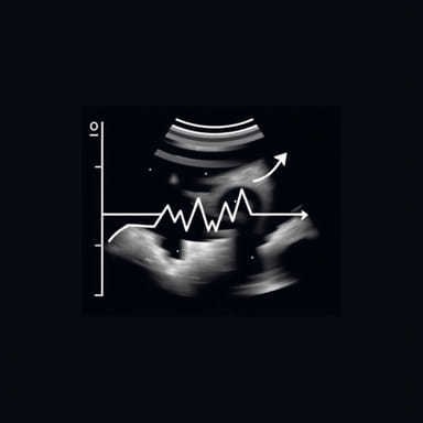
In a healthy, viable intrauterine pregnancy, beta hCG levels typically double every 48 to 72 hours during the first few weeks. This predictable rise helps clinicians estimate gestational age, assess fetal viability, and detect abnormal pregnancy patterns. However, variations can occur, and serial measurements are often more reliable than a single value.
- Rapid rise in early pregnancy suggests normal embryonic development.
- Slow or plateaued levels may indicate a failing pregnancy or ectopic implantation.
- Extremely high values may be associated with multiple gestations or molar pregnancies.
Role of Ultrasound in Early Pregnancy Assessment
Ultrasound imaging is the gold standard for visualizing pregnancy structures. It can confirm the presence of a gestational sac, yolk sac, fetal pole, and heartbeat, which are key markers of viability. The timing of these findings is closely related to beta hCG levels, and their correlation helps determine whether a pregnancy is progressing normally.
Types of Ultrasound Used
- Transvaginal ultrasound– Offers high-resolution imaging, especially useful in early pregnancy (before 8 weeks).
- Transabdominal ultrasound– More common in later pregnancy but can be used once beta hCG levels are high enough for structures to be visible.
Correlation Between Beta hCG and Ultrasound Findings
One of the most clinically important concepts is thediscriminatory zone the beta hCG level above which an intrauterine gestational sac should be visible on ultrasound. For transvaginal ultrasound, this threshold is usually around 1,500 to 2,000 mIU/mL, while for transabdominal scans, it may be 5,000 to 6,500 mIU/mL. If hCG levels exceed the discriminatory zone and no intrauterine sac is seen, the suspicion for ectopic pregnancy rises significantly.
Examples of Correlation
- Beta hCG < 1,000 mIU/mLLikely too early to visualize a gestational sac even with transvaginal ultrasound.
- Beta hCG 1,500-2,000 mIU/mLGestational sac should be visible via transvaginal ultrasound if pregnancy is intrauterine.
- Beta hCG > 6,500 mIU/mLGestational sac should be visible via transabdominal ultrasound.
Clinical Applications of Beta hCG-Ultrasound Correlation
1. Confirming Intrauterine Pregnancy
By comparing beta hCG levels with ultrasound findings, clinicians can determine whether the pregnancy is located within the uterus. This is especially critical in ruling out ectopic pregnancies, which can be life-threatening if undetected.
2. Evaluating Pregnancy Viability
A rising beta hCG accompanied by progressive ultrasound findings (yolk sac, fetal pole, heartbeat) indicates a healthy pregnancy. Conversely, low or plateaued hCG levels combined with absent or regressing ultrasound features may signal miscarriage or anembryonic gestation.
3. Diagnosing Ectopic Pregnancy
If beta hCG levels are above the discriminatory zone but no intrauterine pregnancy is seen, ectopic pregnancy must be strongly considered. Additional evaluation, such as repeat hCG measurements and follow-up ultrasounds, is essential for diagnosis and management.
4. Monitoring High-Risk Pregnancies
Women with a history of recurrent pregnancy loss, tubal surgery, or fertility treatments may require close monitoring using both beta hCG trends and serial ultrasounds. The combined approach provides early detection of complications.
Limitations of Correlation
While the relationship between beta hCG levels and ultrasound findings is a powerful diagnostic tool, it is not without limitations. Biological variability, laboratory differences in hCG assays, and operator-dependent factors in ultrasound interpretation can all affect accuracy.
- Some normal pregnancies may not follow the textbook doubling time for beta hCG.
- Ultrasound visibility depends on equipment quality and operator skill.
- The discriminatory zone is an approximate guide, not an absolute rule.
Best Practices for Clinical Use
To maximize the accuracy of pregnancy assessment using beta hCG and ultrasound correlation, the following steps are recommended
- Use serial beta hCG measurements rather than relying on a single reading.
- Perform ultrasound at appropriate hCG levels to avoid false-negative findings.
- Interpret results in the context of the patient’s symptoms and clinical history.
- Communicate findings clearly to the patient, explaining the need for follow-up if results are inconclusive.
The correlation between beta hCG levels and ultrasound findings is an essential aspect of early pregnancy evaluation. By understanding the expected patterns of hormone rise and the timing of ultrasound visibility, healthcare providers can diagnose pregnancy location and viability more accurately. While no single test is infallible, combining these diagnostic tools offers the best chance for early and accurate assessment, enabling timely intervention and better outcomes for both the patient and the developing pregnancy.