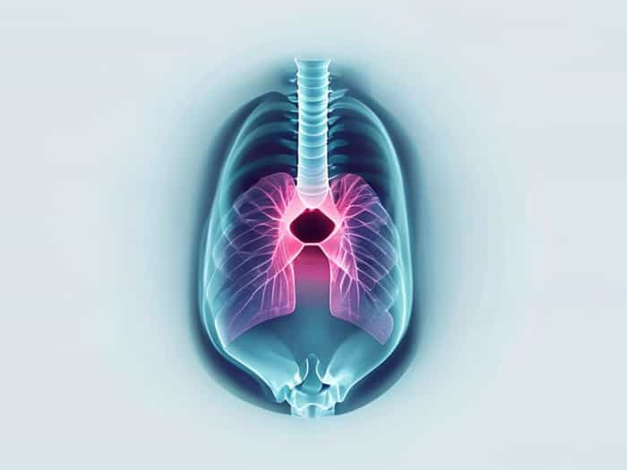
The glottis is a critical anatomical structure located within the larynx, playing an essential role in respiration, phonation, and airway protection. Understanding how the glottis appears on X-ray imaging is vital for medical professionals, particularly radiologists, ENT specialists, and emergency physicians. X-ray evaluation of the glottis can provide valuable information regarding structural abnormalities, injuries, infections, or tumors. While imaging the glottis can be challenging due to its small size and dynamic function, advancements in radiographic techniques have improved visualization, making X-ray a useful tool in diagnosing conditions affecting this crucial part of the airway.
Anatomy of the Glottis
The glottis consists of the vocal cords (true vocal folds) and the space between them, known as the rima glottidis. It is situated within the larynx, above the trachea and below the epiglottis. The glottis serves multiple functions it controls airflow during breathing, produces sound by vibrating the vocal cords, and protects the lower respiratory tract by closing during swallowing. The structural components of the glottis, including the vocal folds, vestibular folds, and surrounding cartilage, all contribute to its function and appearance on imaging studies.
Key Anatomical Features Relevant to X-ray
- True vocal cords responsible for phonation and visible as paired structures on certain radiographic angles.
- Rima glottidis the space between the vocal cords, which can change shape and width depending on breathing and vocalization.
- Epiglottis lies above the glottis and can affect the visualization of the glottal opening.
- Thyroid and cricoid cartilages provide structural landmarks on lateral neck X-rays.
Techniques for Visualizing the Glottis on X-ray
Imaging the glottis with standard X-rays can be challenging due to overlapping structures and its small size. Certain techniques improve the visualization and provide more diagnostic information. Lateral neck X-rays are commonly used to assess the glottis, particularly in cases of suspected airway obstruction, laryngeal injury, or congenital anomalies in children.
Lateral Neck X-ray
A lateral neck X-ray captures the profile of the larynx and glottis, allowing clinicians to assess airway patency, the position of the vocal cords, and any abnormal masses or swelling. Proper positioning is crucial; the patient is typically instructed to extend the neck slightly to optimize the view of the glottis. In adults, the glottis may be less easily distinguished due to the small size and surrounding soft tissue, while in pediatric patients, lateral X-rays can be particularly informative for conditions like croup or epiglottitis.
Other Imaging Considerations
- Dynamic fluoroscopy can be used to observe vocal cord motion during respiration and phonation.
- Contrast studies, though less common, can enhance the visualization of the glottis in complex cases.
- Digital radiography provides improved resolution and allows subtle details of the glottal space to be examined.
Clinical Indications for Glottis X-ray
X-ray imaging of the glottis is often performed when there are concerns about airway obstruction, trauma, infection, or tumors. Early identification of abnormalities in the glottis can prevent severe complications such as respiratory distress or voice impairment. Clinical signs prompting X-ray evaluation may include stridor, hoarseness, difficulty breathing, or throat pain.
Common Conditions Evaluated
- Glottic tumors X-ray can reveal masses, although CT and MRI are often required for detailed assessment.
- Inflammatory conditions such as epiglottitis or laryngitis, which may cause swelling around the glottis.
- Trauma fractures of laryngeal cartilages or soft tissue injury affecting glottal patency.
- Congenital anomalies in pediatric patients, X-ray may detect laryngeal webs, subglottic stenosis, or other structural defects.
Interpreting X-ray Findings of the Glottis
When analyzing X-rays of the glottis, radiologists focus on the width of the rima glottidis, symmetry of the vocal cords, and the presence of any soft tissue abnormalities. Normal glottic appearance shows clear, symmetrical vocal cords with an unobstructed airway. Variations in size, shape, or density may indicate pathology. Careful comparison with clinical findings and, when necessary, supplementary imaging is essential for accurate diagnosis.
Radiographic Signs to Note
- Narrowing of the glottal space may indicate obstruction, inflammation, or mass effect.
- Asymmetry of the vocal cords could suggest paralysis, mass, or scarring.
- Soft tissue swelling often associated with infection or trauma.
- Calcifications in laryngeal cartilage occasionally seen in adults and usually benign.
Limitations of X-ray Imaging
While X-ray can provide useful information, it has limitations in glottis evaluation. The two-dimensional nature of X-rays and overlapping structures can obscure small lesions or subtle functional abnormalities. Additionally, X-rays do not provide the level of detail available with endoscopy, CT, or MRI, which are often required for comprehensive assessment. X-ray should therefore be considered an initial, accessible tool rather than a definitive diagnostic method for glottis pathology.
When to Use Advanced Imaging
- Persistent hoarseness or voice changes that X-ray cannot fully explain.
- Suspected laryngeal tumors or deep tissue involvement.
- Detailed pre-surgical planning for laryngeal or airway procedures.
- Complex trauma involving multiple structures in the neck.
Understanding the glottis on X-ray is a valuable skill for healthcare professionals dealing with airway and laryngeal conditions. While visualization can be challenging, lateral neck X-rays and modern digital techniques provide essential information about the structure and function of the glottis. X-ray imaging is particularly useful for initial evaluation of trauma, obstruction, infection, or congenital anomalies. By combining careful interpretation with clinical findings and supplementary imaging when necessary, medical professionals can effectively assess glottis health and guide appropriate treatment to ensure airway safety and optimal vocal function.