Polymyalgia rheumatica (PMR) is an inflammatory disorder that primarily affects adults over the age of 50, causing pain and stiffness in the shoulders, hips, neck, and proximal limbs. While the diagnosis is largely clinical, imaging studies, including X-rays, can play a supportive role in evaluating patients and excluding other conditions that may mimic PMR. X-ray findings in polymyalgia rheumatica are often subtle or even normal, but understanding their significance, indications, and limitations can help clinicians provide accurate diagnoses and guide management strategies.
Understanding Polymyalgia Rheumatica
Polymyalgia rheumatica is characterized by systemic inflammation, leading to muscle pain and stiffness, particularly in the morning or after periods of inactivity. Although PMR primarily affects the musculoskeletal system, it is closely associated with giant cell arteritis (GCA), which can have serious vascular complications. Laboratory findings often show elevated inflammatory markers, such as erythrocyte sedimentation rate (ESR) and C-reactive protein (CRP). While these findings support the diagnosis, imaging studies, including X-rays, can help rule out other causes of pain, such as osteoarthritis, fractures, or inflammatory arthropathies.
Role of X-ray in Polymyalgia Rheumatica
Plain radiography, or X-ray, is a readily available and cost-effective imaging modality that can be used in patients with suspected polymyalgia rheumatica. While it does not directly visualize inflammation of muscles or bursae, X-rays can identify structural abnormalities that might mimic PMR symptoms. By excluding other pathologies, such as degenerative joint disease, crystal arthropathies, or malignancy, X-rays contribute to a more accurate diagnosis and prevent unnecessary treatments.
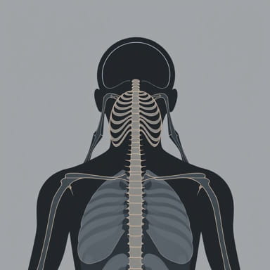
Common Findings on X-ray
In patients with polymyalgia rheumatica, X-ray findings are often subtle. Common observations may include
- Normal joint spaces, as PMR primarily affects soft tissues rather than cartilage.
- Mild osteopenia in proximal long bones due to disuse or chronic inflammation.
- Absence of erosions, which helps differentiate PMR from rheumatoid arthritis.
- Occasional evidence of osteoarthritis or degenerative changes unrelated to PMR.
It is important to note that the lack of significant radiographic abnormalities does not rule out PMR. The disease primarily involves inflammation of periarticular structures, such as bursae, tendons, and synovium, which are not directly visualized on X-ray.
Advanced Imaging Techniques
Although plain X-rays provide limited information in polymyalgia rheumatica, other imaging modalities may be more sensitive in detecting inflammatory changes. These include ultrasound, magnetic resonance imaging (MRI), and positron emission tomography (PET). Ultrasound can detect bursitis, synovitis, and tenosynovitis in the shoulders and hips, which are hallmark features of PMR. MRI provides detailed visualization of soft tissues and can confirm inflammation when the diagnosis is uncertain. PET scans may also reveal systemic inflammation, including vascular involvement suggestive of associated giant cell arteritis.
Indications for X-ray in PMR
Despite its limited sensitivity for detecting inflammation, X-ray remains useful in certain clinical scenarios
- Initial evaluation of patients with shoulder or hip pain to exclude fractures or degenerative joint disease.
- Assessment of chronic pain to rule out osteoarthritis or other structural abnormalities.
- Follow-up imaging to monitor for complications or coexisting musculoskeletal disorders.
- Investigation of atypical presentations or persistent pain not responding to standard therapy.
Differential Diagnosis Using X-ray
One of the key roles of X-ray in polymyalgia rheumatica is aiding differential diagnosis. Since PMR symptoms overlap with other musculoskeletal and rheumatologic disorders, radiographic evaluation helps differentiate between conditions such as
- Rheumatoid arthritis, which typically shows joint erosions and narrowing of joint spaces.
- Osteoarthritis, characterized by osteophyte formation, joint space narrowing, and subchondral sclerosis.
- Rotator cuff disease or bursitis, which may show secondary bone changes in chronic cases.
- Metastatic bone disease, which may present as lytic or sclerotic lesions.
Clinical Integration of X-ray Findings
While X-rays rarely provide definitive evidence of polymyalgia rheumatica, their role in excluding other conditions makes them valuable in clinical practice. When combined with clinical evaluation, laboratory tests, and patient history, X-ray findings help clinicians make more informed decisions. For example, the absence of erosions or significant joint space loss in a patient with proximal limb pain and elevated ESR supports the diagnosis of PMR over rheumatoid arthritis or degenerative joint disease.
Limitations of X-ray in PMR
Plain radiography has notable limitations in the context of polymyalgia rheumatica
- It cannot directly visualize soft tissue inflammation, such as bursitis or synovitis.
- Early disease may appear entirely normal, leading to potential diagnostic uncertainty.
- Subtle structural changes may be overlooked, particularly in small joints or periarticular regions.
- It is less useful for monitoring disease activity or response to therapy.
Management and Follow-Up
Management of polymyalgia rheumatica typically involves corticosteroid therapy to reduce inflammation and relieve symptoms. While X-rays are not used to monitor disease activity, they can be valuable in follow-up when patients develop new symptoms or complications. Clinicians may order repeat imaging to assess for coexisting osteoarthritis, fractures, or other structural changes that could influence ongoing treatment.
Polymyalgia rheumatica X-ray findings are often subtle or even normal, reflecting the primarily inflammatory and periarticular nature of the disease. However, X-rays remain an important tool for excluding other musculoskeletal disorders and guiding the diagnostic process. By providing a cost-effective and readily available method to evaluate structural abnormalities, plain radiography supports the clinical management of PMR. When combined with patient history, physical examination, and laboratory data, X-ray helps clinicians distinguish PMR from conditions with overlapping symptoms, ensuring accurate diagnosis and effective treatment. Advanced imaging techniques, such as ultrasound or MRI, may complement X-ray findings, offering more detailed assessment of soft tissue inflammation and improving diagnostic confidence.