The lungs are essential organs of respiration, located within the thoracic cavity and protected by the rib cage. They play a vital role in the exchange of oxygen and carbon dioxide, making them critical for sustaining life. To understand their location, boundaries, and relation to other structures, it is important to learn about the surface marking of the lung. Surface markings help medical students, healthcare professionals, and surgeons visualize the position of the lungs and their parts during examination, diagnosis, and surgery, without directly viewing the organs inside the body.
Overview of Lung Surface Marking
The surface marking of the lung refers to the anatomical landmarks that indicate the outlines of the lungs and their fissures on the chest wall. These markings correspond to the pleural reflections and help identify where each part of the lung lies relative to the ribs and other structures. Since the lungs are not symmetrical the right lung has three lobes, while the left lung has two their surface markings differ slightly. Understanding these differences is essential for interpreting imaging studies, performing physical examinations, and carrying out certain clinical procedures like thoracentesis.
General Anatomy of the Lungs
Before discussing surface markings, it is helpful to recall the basic structure of the lungs
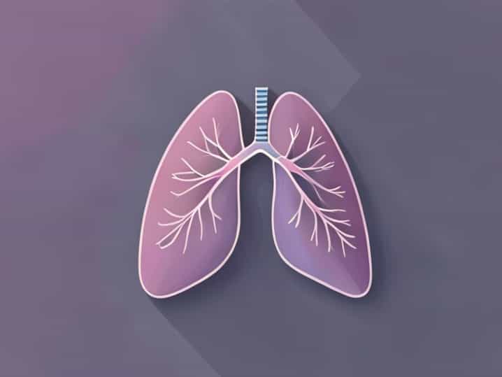
- Right LungDivided into three lobes upper (superior), middle, and lower (inferior) separated by horizontal and oblique fissures.
- Left LungDivided into two lobes upper and lower separated by a single oblique fissure. It also has a cardiac notch and a lingula, which accommodate the heart.
- ApexThe topmost portion of each lung, which extends above the level of the first rib into the root of the neck.
- BaseThe bottom portion resting on the diaphragm.
- SurfacesEach lung has three surfaces costal (facing the ribs), mediastinal (facing the heart and great vessels), and diaphragmatic (facing downward).
These structural details form the foundation for understanding where the lungs can be palpated, auscultated, or visualized on the chest wall.
Surface Marking of the Right Lung
The right lung’s surface markings can be traced along the chest wall using anatomical landmarks such as ribs, costal cartilages, and intercostal spaces. Below is a simplified outline of the right lung’s surface boundaries and fissures.
Upper Border
The apex of the right lung extends about 2.5 cm (1 inch) above the medial third of the clavicle, behind the sternoclavicular joint. From this point, the line descends behind the sternoclavicular joint to meet the second costal cartilage next to the sternum. This indicates the beginning of the anterior border.
Anterior Border
The anterior border runs vertically downward from the sternal end of the second costal cartilage to the sixth costal cartilage close to the midline. The border separates the costal and mediastinal surfaces. The right lung’s anterior margin is straight because it does not have a cardiac notch like the left lung.
Lower Border
The lower border begins at the sixth costal cartilage near the sternum and runs laterally, crossing the eighth rib at the midaxillary line and the tenth rib at the posterior axillary line. Posteriorly, it ends at the level of the tenth thoracic vertebra. This line represents the base of the lung resting on the diaphragm.
Posterior Border
The posterior border extends from the apex down to the level of the tenth thoracic vertebra. It lies about 1.5 cm lateral to the spinous processes and marks the posterior limit of lung tissue in contact with the chest wall.
Fissures of the Right Lung
- Oblique FissureStarts from the level of the spine of the T3 vertebra, runs downward and forward across the posterior chest wall, and ends at the sixth costal cartilage near the midclavicular line. It separates the lower lobe from the upper and middle lobes.
- Horizontal FissureRuns horizontally from the oblique fissure along the level of the fourth costal cartilage to the sternum. It separates the upper and middle lobes.
These fissures are significant during auscultation and imaging, as they help locate specific lobes where disease or fluid might accumulate.
Surface Marking of the Left Lung
The left lung, although similar in structure, differs in a few key ways because it has only two lobes and must accommodate the heart. Its surface marking lines are therefore slightly different.
Upper Border
The apex of the left lung also extends about 2.5 cm above the medial third of the clavicle. From this point, the border runs downward behind the sternoclavicular joint and meets the second costal cartilage near the sternum, just like the right lung.
Anterior Border
The anterior border of the left lung descends to the level of the fourth costal cartilage. At this level, it curves laterally and downward, forming thecardiac notch. The notch allows space for the heart, particularly the left ventricle, creating an indentation on the medial surface of the lung. Below the notch, the border continues downward to the sixth costal cartilage at the midclavicular line.
Lower Border
The lower border follows a similar pattern to the right lung, extending from the sixth costal cartilage at the midclavicular line to the eighth rib in the midaxillary line, and to the tenth rib posteriorly. It also reaches the level of the tenth thoracic vertebra at the back.
Posterior Border
The posterior border extends from the apex down to the level of the T10 vertebra. It is slightly curved and corresponds to the line where the lung meets the vertebral column.
Fissure of the Left Lung
- Oblique FissureThis is the only fissure in the left lung. It starts at the level of the spine of the T3 vertebra, passes obliquely downward across the back, and ends near the sixth costal cartilage anteriorly. It separates the upper lobe from the lower lobe.
Pleural Reflections and Relation to Lung Markings
The pleura is a double-layered membrane surrounding each lung. Theparietal pleuralines the thoracic cavity, while thevisceral pleuracovers the lung itself. The space between them is the pleural cavity. The surface markings of the pleura extend slightly below those of the lungs, providing a potential space where fluid can collect in conditions like pleural effusion.
For example, the pleural reflection reaches the eighth rib in the midclavicular line, the tenth rib in the midaxillary line, and the twelfth rib posteriorly. This difference between lung and pleura boundaries is clinically important when inserting a needle to remove pleural fluid the procedure must stay below the lung margin but within the pleural space.
Clinical Significance of Lung Surface Markings
Knowledge of lung surface markings is critical in medical practice. It allows healthcare professionals to identify areas of the lung during physical examination, radiography, and surgical procedures. For example
- Percussion and AuscultationPhysicians use surface markings to determine which lobe they are listening to when checking for breath sounds.
- ThoracentesisThe surface markings help identify safe sites for inserting a needle into the pleural space, typically above the eighth rib in the midaxillary line.
- RadiologyInterpreting chest X-rays or CT scans requires understanding how the lobes and fissures of the lungs are arranged beneath the ribs and sternum.
- Surgical ProceduresDuring lung resections or biopsies, precise knowledge of lung anatomy ensures minimal damage to surrounding structures.
Examples in Clinical Context
When a physician detects decreased breath sounds or dullness to percussion in the lower part of the chest, they can use lung surface markings to localize the problem. If the dullness is below the level of the tenth rib in the midaxillary line, it may indicate pleural effusion rather than lung pathology. Similarly, understanding the location of fissures helps identify whether pneumonia or fluid is confined to a single lobe or has spread more widely.
Surface Markings in Radiological Correlation
Modern imaging techniques like X-rays and CT scans visualize lung structures in great detail, but anatomical surface marking remains foundational for interpreting them. For example, the oblique fissure corresponds roughly to a line from the level of T3 posteriorly to the sixth costal cartilage anteriorly. Knowing this helps radiologists distinguish between lobes on a frontal or lateral chest X-ray. Similarly, understanding where the pleural reflections lie can help diagnose pneumothorax or effusion.
Summary and Final Thoughts
The surface marking of the lung provides a practical map for identifying the position of the lungs, lobes, and fissures on the chest wall. The right lung has three lobes separated by two fissures, while the left lung has two lobes and one fissure, along with a cardiac notch. These surface landmarks are essential for physical examination, diagnostic imaging, and clinical interventions. By combining anatomical knowledge with careful observation, medical professionals can accurately interpret lung findings, diagnose respiratory conditions, and perform procedures safely. Understanding these surface markings bridges the gap between theory and clinical practice, making it one of the most valuable aspects of applied anatomy in medicine.