Distal femoral cortical irregularity is a term commonly encountered in radiology reports, referring to subtle variations or disruptions in the smooth contour of the distal femur’s cortical bone. The distal femur is the lower portion of the thigh bone that forms part of the knee joint, and cortical irregularities in this region can be incidental findings or indicators of underlying pathology. Radiologists and orthopedic specialists often assess these irregularities in detail to differentiate between normal anatomical variations, developmental changes, and clinically significant conditions. Understanding the radiological features, causes, and implications of distal femoral cortical irregularities is essential for accurate diagnosis and appropriate management.
Understanding Distal Femoral Cortical Irregularity
The cortex of a bone is its dense outer layer, providing structural support and protection. In radiological imaging, cortical irregularity refers to changes in the continuity, thickness, or smoothness of this outer layer. At the distal femur, irregularities may appear as small notches, undulations, or areas of thinning. These findings are often subtle and may require high-resolution imaging techniques, such as X-ray, computed tomography (CT), or magnetic resonance imaging (MRI), to evaluate accurately.
Radiological Appearance
- X-rayCortical irregularities can present as small areas of lucency or focal disruptions along the bone cortex.
- CT scanOffers detailed cross-sectional images, allowing better assessment of the cortical surface and identification of subtle lesions or defects.
- MRIUseful in evaluating associated soft tissue or marrow changes, particularly if an underlying pathology is suspected.
Common Causes of Distal Femoral Cortical Irregularity
Distal femoral cortical irregularities may arise from a variety of causes, ranging from benign anatomical variations to pathological conditions. Correct interpretation is crucial to avoid unnecessary interventions or misdiagnosis.
Normal Anatomical Variants
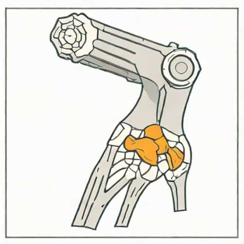
Some cortical irregularities are simply normal variants. The distal femur has areas where tendons and ligaments attach, such as the adductor tubercle and epicondyles. These insertion sites can create small notches or irregularities that appear on imaging but are not clinically significant. Recognizing these variants helps prevent misinterpretation as fractures or tumors.
Developmental Changes
In younger individuals, cortical irregularities may result from ongoing bone growth and remodeling. The distal femur continues to develop into late adolescence, and irregularities can reflect normal physeal (growth plate) activity or minor areas of cortical remodeling. Radiologists must consider patient age and skeletal maturity when evaluating these findings.
Trauma and Stress Injuries
Microfractures or stress-related injuries can also cause cortical irregularity. Repetitive mechanical stress, such as in athletes or physically active individuals, may lead to small cortical defects known as stress reactions or stress fractures. These are often associated with localized pain and may progress if not managed appropriately.
Pathological Conditions
While many cortical irregularities are benign, some may indicate underlying pathology. Possible conditions include
- OsteomyelitisInfection of the bone can cause cortical destruction and irregularity, often accompanied by soft tissue swelling and systemic symptoms.
- Bone tumorsBenign tumors like osteochondromas or fibrous cortical defects may manifest as cortical irregularities. Malignant lesions, although rarer, can also disrupt the cortical surface.
- Osteoporosis or metabolic bone diseaseReduced bone density can make the cortex more susceptible to irregularities or microfractures.
Clinical Significance
Identifying the cause of distal femoral cortical irregularity is essential to determine whether intervention is needed. Many irregularities are incidental findings with no clinical impact, especially when associated with normal anatomical landmarks or growth-related changes. However, irregularities resulting from trauma, infection, or tumors require prompt evaluation and management.
Symptoms and Evaluation
Clinical presentation can vary depending on the underlying cause
- Pain or tenderness localized to the distal femur, particularly after activity or trauma.
- Swelling, redness, or warmth if infection or inflammatory conditions are present.
- Functional limitations such as difficulty walking, bending the knee, or bearing weight.
Radiological findings are often correlated with patient history, physical examination, and sometimes laboratory tests. For example, an irregularity associated with elevated inflammatory markers may suggest osteomyelitis, whereas an asymptomatic cortical defect in a young adult is more likely a benign variant.
Management Strategies
Management of distal femoral cortical irregularities depends on their etiology. In many cases, observation and follow-up imaging are sufficient, particularly when the irregularity is deemed a normal variant or developmental change. Specific interventions may include
- Conservative managementRest, activity modification, and pain management for stress-related cortical defects or minor trauma.
- Antibiotic therapyRequired for osteomyelitis or infectious causes, often combined with immobilization or surgical drainage in severe cases.
- Surgical interventionIndicated for symptomatic benign tumors, pathological fractures, or malignant lesions.
- MonitoringRegular imaging follow-up to track changes in cortical irregularities over time.
Importance of Accurate Radiological Interpretation
Radiologists play a crucial role in distinguishing between benign and pathological distal femoral cortical irregularities. Accurate interpretation prevents unnecessary procedures and ensures appropriate care. Factors considered include the shape, location, size, and margins of the irregularity, as well as associated findings in the surrounding bone and soft tissue. Cross-sectional imaging such as CT or MRI is often employed when initial X-rays are inconclusive.
Distal femoral cortical irregularity is a radiological finding that can range from a normal anatomical variant to an indicator of serious pathology. Understanding the mechanisms behind these irregularities, including developmental, traumatic, and pathological causes, is essential for proper diagnosis and management. Careful correlation of imaging findings with clinical symptoms and patient history allows for accurate assessment, minimizing unnecessary interventions while ensuring timely treatment for conditions that require attention. Awareness of these factors helps healthcare professionals provide optimal care and improves patient outcomes when distal femoral cortical irregularities are identified on radiology reports.