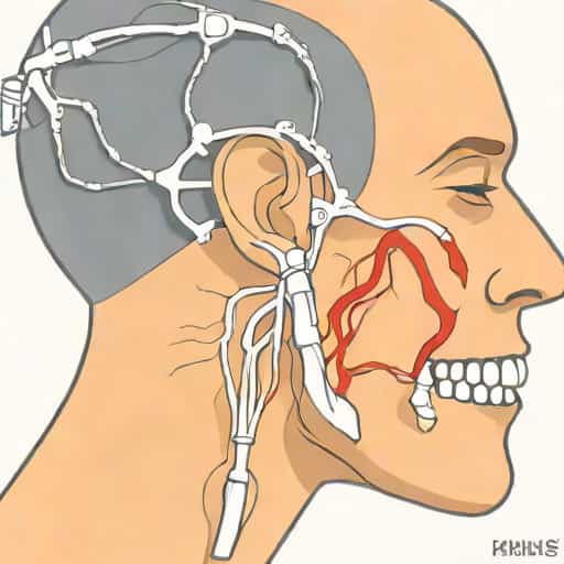
The maxillary nerve, also known as the second branch of the trigeminal nerve (cranial nerve V2), is a crucial component of the human nervous system that plays an essential role in facial sensation. It provides sensory innervation to the midfacial region, including the upper jaw, teeth, nasal cavity, palate, and parts of the face and sinuses. Understanding the maxillary nerve is important for students of anatomy, dentistry, neurology, and surgery because it helps explain the pathways of sensation, pain, and reflexes in the face. Kenhub, a renowned educational platform, provides detailed resources on the maxillary nerve, highlighting its anatomical course, branches, clinical significance, and related pathologies. This topic explores the anatomy of the maxillary nerve, its function, clinical applications, and key insights provided by Kenhub to give a thorough understanding of this essential nerve.
Anatomy of the Maxillary Nerve
The maxillary nerve is one of three main branches of the trigeminal nerve, the largest cranial nerve responsible for sensory and motor functions in the face. Unlike the mandibular nerve, which has both motor and sensory functions, the maxillary nerve is purely sensory. It originates from the trigeminal ganglion, located in Meckel’s cave within the cranial cavity, and exits the skull through the foramen rotundum into the pterygopalatine fossa. From there, it branches extensively to supply sensation to the midface.
Course of the Maxillary Nerve
The pathway of the maxillary nerve begins at the trigeminal ganglion and passes through the foramen rotundum. It enters the pterygopalatine fossa, a small space behind the maxilla that houses the pterygopalatine ganglion. Within this region, the maxillary nerve gives off several branches, including the zygomatic, infraorbital, and posterior superior alveolar nerves. These branches extend to the face, oral cavity, and nasal structures, allowing the nerve to provide comprehensive sensory coverage of the midfacial region.
Key Branches of the Maxillary Nerve
- Zygomatic nerveSupplies the skin over the zygomatic and temporal regions.
- Infraorbital nerveContinues from the maxillary nerve through the infraorbital foramen, providing sensation to the lower eyelid, upper lip, and lateral nose.
- Posterior superior alveolar nerveInnervates the molar teeth and associated gingiva.
- Palatine nervesInclude the greater and lesser palatine nerves, supplying the hard and soft palate.
- Nasal branchesProvide sensation to the nasal cavity and septum.
Function of the Maxillary Nerve
The maxillary nerve is primarily responsible for conveying sensory information from the midface to the central nervous system. This includes touch, pain, temperature, and proprioception. Its sensory distribution is essential for detecting stimuli from the upper jaw, upper teeth, maxillary sinuses, nasal cavity, palate, and areas of the face around the cheeks and lower eyelids. Through these sensory pathways, the maxillary nerve plays a critical role in reflexes, facial sensation, and protective mechanisms.
Sensory Areas
The sensory areas supplied by the maxillary nerve can be grouped as follows
- Midfacial skin, including the cheeks, lower eyelids, and upper lip.
- Upper teeth and gums, particularly the premolars and molars.
- Maxillary sinus and nasal cavity structures.
- Palate, including the hard and soft palate.
Clinical Significance
Understanding the maxillary nerve has significant implications in medicine and dentistry. Damage or irritation of this nerve can result in various sensory disturbances, pain syndromes, and complications during surgical procedures. Clinicians rely on anatomical knowledge of the maxillary nerve for accurate diagnosis, anesthesia administration, and surgical planning.
Trigeminal Neuralgia
Trigeminal neuralgia is a condition characterized by sudden, severe facial pain along the distribution of the trigeminal nerve, including the maxillary branch. Patients may experience sharp, stabbing pain triggered by touch, chewing, or even speaking. Identifying the specific branch affected, such as the maxillary nerve, is essential for targeted treatment and management.
Dental Procedures and Anesthesia
The maxillary nerve is particularly relevant in dentistry. Local anesthetic blocks targeting branches like the posterior superior alveolar or infraorbital nerves are commonly used to numb the upper teeth and midface during procedures such as tooth extractions, root canals, and restorative work. Accurate knowledge of the nerve’s course ensures effective anesthesia and minimizes patient discomfort.
Sinus and Facial Surgery
Surgeries involving the maxillary sinus, orbital floor, or midfacial region require careful consideration of the maxillary nerve to avoid sensory deficits. Injury to the nerve during surgical intervention can result in numbness, tingling, or chronic pain, highlighting the importance of precise anatomical understanding.
Kenhub Resources on the Maxillary Nerve
Kenhub provides comprehensive educational materials that enhance the understanding of the maxillary nerve. Through detailed anatomical diagrams, interactive learning tools, and clinical correlation explanations, Kenhub helps students and professionals grasp the nerve’s course, branches, and function. Key features of Kenhub resources include
Visual Guides and Anatomy Charts
Kenhub offers high-quality illustrations of the trigeminal nerve, pterygopalatine fossa, and maxillary nerve branches. These visuals help learners identify structures in relation to the skull, oral cavity, and facial landmarks, making it easier to memorize anatomical relationships.
Clinical Correlations
Kenhub integrates clinical scenarios, including dental anesthesia, trigeminal neuralgia, and maxillofacial surgery, to show the practical applications of the maxillary nerve. This approach bridges the gap between theoretical knowledge and real-world medical practice.
Interactive Learning Tools
Interactive quizzes, 3D models, and labeled diagrams allow users to test their understanding of the maxillary nerve. These tools promote active learning and retention, making complex anatomy more approachable for students and professionals.
Tips for Studying the Maxillary Nerve
For those seeking to master the anatomy and function of the maxillary nerve, several study strategies can enhance learning outcomes. Utilizing multiple resources, engaging in active recall, and applying clinical examples can reinforce understanding and application.
- Review diagrams of the trigeminal nerve and its branches regularly.
- Use mnemonics to remember key branches and their sensory areas.
- Practice labeling exercises to improve recall of anatomical structures.
- Relate anatomical knowledge to clinical scenarios, such as dental anesthesia or neuralgia.
- Use interactive tools and 3D models to visualize spatial relationships.
The maxillary nerve, as the second branch of the trigeminal nerve, plays a vital role in facial sensation, dental procedures, and clinical diagnosis. Its complex anatomy, extensive branching, and sensory distribution make it a critical structure in medicine and dentistry. Resources from Kenhub provide valuable guidance for understanding the maxillary nerve, offering visual, interactive, and clinically relevant materials. By mastering its anatomy, function, and clinical significance, students and professionals can apply their knowledge effectively in both academic and practical settings. Awareness of the maxillary nerve is essential not only for learning anatomy but also for ensuring patient safety and optimal outcomes in medical, dental, and surgical interventions.