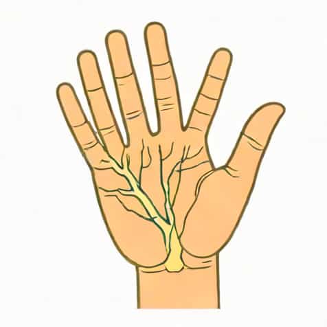
The ulnar nerve is one of the major nerves of the upper limb, playing a crucial role in both motor and sensory functions. Its sensory supply is of particular importance for clinicians, physiotherapists, and anatomy students, as it helps in diagnosing nerve injuries, understanding hand sensation, and guiding treatment plans. The ulnar nerve originates from the medial cord of the brachial plexus, carrying fibers primarily from the C8 and T1 spinal nerve roots. It travels down the arm, behind the medial epicondyle of the humerus, and continues along the forearm to the hand. The sensory distribution of the ulnar nerve is distinctive, covering specific areas of the hand and forearm, which are critical in assessing nerve integrity and understanding neuropathic conditions.
Anatomy of the Ulnar Nerve
The ulnar nerve arises from the medial cord of the brachial plexus, containing fibers from the C8 and T1 nerve roots. It descends along the medial aspect of the arm, running adjacent to the brachial artery, before passing posterior to the medial epicondyle at the elbow-a location commonly referred to as the funny bone due to its susceptibility to compression or trauma. After crossing the elbow, the nerve enters the forearm, traveling between the flexor carpi ulnaris and flexor digitorum profundus muscles, providing motor innervation along the way. Its sensory branches extend to the skin of the forearm, hand, and fingers, forming the basis of its sensory supply.
Course of the Ulnar Nerve
- Originates from the medial cord of the brachial plexus (C8-T1 fibers).
- Descends along the medial arm, near the brachial artery.
- Passes posterior to the medial epicondyle of the humerus.
- Enters the forearm between flexor carpi ulnaris and flexor digitorum profundus muscles.
- Continues into the hand via Guyon’s canal, dividing into superficial and deep branches.
Sensory Distribution of the Ulnar Nerve
The ulnar nerve provides sensory innervation to both the forearm and hand, supplying specific areas that allow clinicians to test nerve function effectively. In the forearm, a branch known as the dorsal cutaneous branch emerges approximately 5-7 cm proximal to the wrist, providing sensation to the dorsal medial hand and dorsal surfaces of the little finger and half of the ring finger. In the hand, the ulnar nerve divides into superficial and deep branches. The superficial branch primarily carries sensory fibers, innervating the palmar aspect of the little finger and half of the ring finger, as well as the associated area of the palm. This precise distribution is essential for detecting ulnar nerve lesions and distinguishing them from other peripheral nerve injuries.
Dorsal Cutaneous Branch
- Branches off the ulnar nerve in the distal forearm.
- Supplies sensation to the dorsal medial hand.
- Innervates the dorsal surfaces of the little finger and medial half of the ring finger.
Superficial Branch of the Ulnar Nerve
- Arises after the ulnar nerve enters the hand through Guyon’s canal.
- Provides sensory innervation to the palmar surface of the little finger and medial half of the ring finger.
- Supplies the hypothenar eminence and adjacent palm area.
Deep Branch (Primarily Motor but Minor Sensory Contribution)
While the deep branch of the ulnar nerve is mainly responsible for motor innervation to intrinsic hand muscles, it may carry minor sensory fibers to periarticular structures and contribute to proprioceptive feedback. Its role in sensory supply is limited but significant in complex clinical assessments.
Clinical Significance of Ulnar Nerve Sensory Supply
The sensory distribution of the ulnar nerve is clinically significant for diagnosing nerve injuries, entrapment syndromes, and neuropathies. Compression at the elbow, often due to prolonged pressure or trauma, can result in sensory loss along the dorsal and palmar surfaces of the medial hand. Similarly, compression in Guyon’s canal at the wrist, commonly seen in cyclists or individuals with repetitive hand activities, can produce numbness, tingling, or sensory deficits in the ulnar-innervated fingers. Clinicians often use sensory tests, including light touch, pinprick, and two-point discrimination, to evaluate ulnar nerve function and determine the site of lesion.
Common Conditions Affecting Ulnar Nerve Sensation
- Cubital tunnel syndrome – compression at the elbow affecting dorsal and palmar sensation.
- Guyon’s canal syndrome – wrist compression leading to sensory loss in the little and ring fingers.
- Traumatic injury – fractures or lacerations causing localized sensory deficits.
- Peripheral neuropathy – systemic conditions like diabetes affecting ulnar nerve fibers.
Testing Ulnar Nerve Sensory Function
Assessing the sensory supply of the ulnar nerve involves careful examination of both dorsal and palmar aspects of the hand. Standard tests include
Light Touch Test
- Gently touch the dorsal and palmar surfaces of the little finger and medial half of the ring finger.
- Compare sensation bilaterally to detect asymmetry.
Pinprick Test
- Use a sterile pin to lightly prick the skin in the ulnar distribution area.
- Observe patient’s perception of sharp versus dull sensations.
Two-Point Discrimination
- Use calipers to test the ability to perceive two separate points on the little and ring fingers.
- Reduced discrimination may indicate ulnar nerve compromise.
Anatomical Variations and Considerations
While the ulnar nerve has a fairly predictable course and sensory distribution, anatomical variations are common. Some individuals may have differences in the branching pattern of the dorsal cutaneous or superficial branches, which can affect sensory presentation. Awareness of these variations is important for surgeons, anesthetists, and clinicians performing nerve blocks, surgical procedures, or diagnostic evaluations. Detailed knowledge of these variations ensures accurate assessment and prevents iatrogenic injury.
Key Variations
- Differences in dorsal cutaneous branch origin and branching pattern.
- Variability in the area of palmar innervation of the superficial branch.
- Occasional overlap with median or radial nerve territories.
The ulnar nerve sensory supply is a vital aspect of upper limb anatomy, providing sensation to specific areas of the forearm and hand. Understanding its course, branches, and clinical significance allows for accurate diagnosis of nerve injuries and effective management of related conditions. The dorsal cutaneous branch supplies the medial dorsal hand and dorsal fingers, while the superficial branch innervates the palmar surfaces of the little and half of the ring finger. Knowledge of these sensory territories is essential for healthcare professionals to assess nerve function, guide treatment, and improve patient outcomes. Awareness of anatomical variations further enhances clinical precision, making mastery of the ulnar nerve sensory supply indispensable in medical practice.