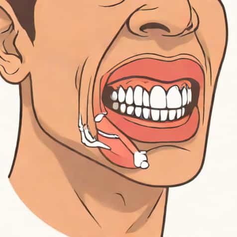
Mastication, commonly known as chewing, is a fundamental process in human digestion that involves the breakdown of food into smaller, more manageable pieces before it reaches the stomach. This process is not only essential for mechanical digestion but also facilitates the chemical digestion of food by increasing its surface area. The main muscles responsible for mastication are highly specialized, allowing precise movements of the jaw and efficient processing of a wide variety of foods. Understanding which muscles are involved, their function, and how they work together provides insight into both human anatomy and the mechanics of eating.
The Primary Muscle of Mastication
The main muscle used for mastication is the masseter muscle. The masseter is a thick, rectangular muscle located in the cheek area, extending from the zygomatic arch (cheekbone) down to the lateral surface of the mandible (lower jaw). It is one of the strongest muscles in the human body relative to its size and plays a central role in elevating the mandible, which closes the mouth and allows for biting and chewing.
Anatomy of the Masseter Muscle
The masseter has two main layers the superficial layer and the deep layer. Each layer contributes to specific movements of the jaw
- Superficial LayerThis layer originates from the anterior portion of the zygomatic arch and inserts into the angle and lower part of the mandible. It is primarily responsible for powerful jaw closure during biting.
- Deep LayerOriginating from the posterior portion of the zygomatic arch and inserting into the upper ramus of the mandible, the deep layer assists in stabilizing the jaw and producing precise grinding movements during chewing.
Together, these layers allow the masseter to generate significant force, enabling humans to chew a variety of foods from soft fruits to tougher meat or nuts.
Other Muscles Involved in Mastication
While the masseter is the primary muscle for chewing, it works in conjunction with other muscles of mastication to ensure smooth and efficient jaw movement. These muscles include the temporalis, medial pterygoid, and lateral pterygoid muscles.
Temporalis Muscle
The temporalis muscle is a fan-shaped muscle located on the side of the head, covering the temporal bone. Its main function is to elevate and retract the mandible, assisting the masseter in closing the mouth. The anterior fibers primarily lift the jaw, while the posterior fibers retract it, allowing for controlled and coordinated chewing movements.
Medial Pterygoid Muscle
The medial pterygoid is a thick muscle found on the inner side of the mandible. It functions in elevating the jaw, similar to the masseter, but also contributes to small side-to-side movements of the jaw, which are important for grinding food between the molars. This muscle works in tandem with the lateral pterygoid to produce smooth lateral motions necessary for efficient mastication.
Lateral Pterygoid Muscle
The lateral pterygoid is unique among the mastication muscles because it primarily functions to open the jaw, protrude the mandible, and enable side-to-side movements. By pulling the mandibular condyle forward, it works against the masseter and temporalis to allow controlled jaw opening, which is essential for taking bites and repositioning food during chewing.
Coordination of Mastication Muscles
Mastication is a complex process that requires precise coordination between the muscles of the jaw, the teeth, and the temporomandibular joint (TMJ). When the masseter contracts to close the jaw, the medial pterygoid supports the upward movement while the temporalis stabilizes and retracts the mandible. Simultaneously, the lateral pterygoid ensures smooth jaw opening when needed. This coordination allows humans to grind, crush, and manipulate food efficiently.
Role of the Temporomandibular Joint
The TMJ acts as a hinge and sliding joint, connecting the mandible to the temporal bone of the skull. The muscles of mastication, particularly the masseter, rely on the TMJ to facilitate complex jaw movements. Proper function of this joint is essential for effective mastication, as dysfunction can lead to pain, difficulty chewing, and reduced bite force.
Importance of the Masseter Muscle in Daily Life
The masseter muscle is not only crucial for chewing but also plays a role in speech, facial expression, and overall oral health. A strong masseter ensures effective grinding of food, which aids digestion and nutrient absorption. Weakness or injury to the masseter can result in difficulty eating, jaw fatigue, or imbalance in other facial muscles.
Clinical Considerations
- Temporomandibular DisordersDysfunction in the masseter or associated muscles can contribute to TMJ disorders, causing pain, clicking, or difficulty opening the mouth.
- BruxismExcessive clenching or grinding of teeth often involves overactivity of the masseter, potentially leading to muscle hypertrophy or jaw discomfort.
- RehabilitationPhysical therapy exercises targeting the masseter and surrounding muscles can improve jaw strength and restore function after injury.
The main muscle used for mastication is the masseter, a powerful and essential muscle responsible for elevating the jaw and enabling effective chewing. While the masseter provides the primary force for biting, it works in coordination with the temporalis, medial pterygoid, and lateral pterygoid muscles to produce precise movements necessary for grinding and processing food. Understanding the anatomy and function of these muscles not only highlights the complexity of human mastication but also emphasizes the importance of jaw health in digestion, speech, and overall well-being. Maintaining strong and functional mastication muscles ensures efficient food breakdown and contributes to oral and digestive health, making the masseter an essential component of daily life.