The pelvic inlet is a critical anatomical landmark that defines the boundary between the abdominal cavity and the true pelvis. It is especially important in obstetrics, anatomy, and surgery because its size and shape influence childbirth, pelvic organ function, and the surgical approach to the pelvis. Understanding which anatomic structures define the pelvic inlet allows medical professionals to assess pelvic dimensions accurately, anticipate complications during labor, and understand the overall pelvic architecture. The pelvic inlet, also called the superior pelvic aperture, is bounded by several bony landmarks that together form a continuous ring, providing structural support and guiding the passage of internal organs.
An Overview of the Pelvic Inlet
The pelvic inlet serves as the upper opening of the true pelvis. It separates the false pelvis, which lies above and supports abdominal organs, from the true pelvis, which contains pelvic organs such as the bladder, rectum, and reproductive structures. The inlet is crucial in obstetrics, as its dimensions determine whether a fetus can pass safely during vaginal delivery. In clinical practice, the pelvic inlet is evaluated by palpation, imaging techniques, and anthropometric measurements to assess pelvic adequacy.
Shape and Clinical Significance
The shape of the pelvic inlet varies between individuals and between sexes. In females, the inlet is typically wider and more oval to facilitate childbirth, while in males, it is narrower and more heart-shaped. Knowledge of these anatomical differences is important in gynecology, obstetrics, and orthopedic surgery. The inlet not only affects the passage of the fetus but also guides the orientation and position of pelvic organs.
Bony Structures Defining the Pelvic Inlet
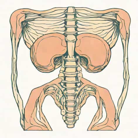
The pelvic inlet is defined by a continuous ring of bony structures that form the margin of the superior pelvic aperture. These structures include the sacral promontory, arcuate lines of the ilia, pectineal lines of the pubic bones, and the upper border of the pubic symphysis. Together, these landmarks form the pelvic brim, which is the anatomical boundary of the pelvic inlet.
Sacral Promontory
The sacral promontory is the anterior projecting edge of the first sacral vertebra. It is a prominent landmark that forms the posterior boundary of the pelvic inlet. Clinically, it can be palpated during pelvic examinations, and it serves as a reference point in obstetric measurements. The sacral promontory contributes to the anteroposterior diameter of the pelvic inlet, which is one of the key dimensions considered during labor.
Arcuate Lines of the Ilium
The arcuate lines are smooth, curved ridges on the internal surface of the ilium. They run from the sacroiliac joints toward the pubic bones and form the lateral margins of the pelvic inlet. These lines help define the lateral boundaries of the pelvic brim and provide attachment points for ligaments and muscles. The arcuate lines are significant in maintaining the structural integrity of the pelvis and supporting pelvic organs.
Pectineal Lines of the Pubis
The pectineal lines, also called the pecten pubis, are ridges along the superior pubic ramus. They form part of the anterior boundary of the pelvic inlet and connect with the arcuate lines laterally. The pectineal lines provide structural reinforcement and serve as landmarks for surgical procedures, including hernia repair and pelvic osteotomies.
Upper Border of the Pubic Symphysis
The pubic symphysis is a cartilaginous joint between the left and right pubic bones. The upper border of this joint completes the anterior margin of the pelvic inlet. It is clinically significant because its position helps define the anterior pelvic diameter. During childbirth, the distance from the sacral promontory to the upper border of the pubic symphysis, called the obstetric conjugate, is a critical measurement for assessing fetal passage.
Pelvic Brim and Its Components
The pelvic brim is the continuous ridge formed by the bony structures of the pelvic inlet. It serves as a structural and functional boundary that distinguishes the true pelvis from the false pelvis. The components of the pelvic brim include
- Posteriorly Sacral promontory
- Laterally Arcuate lines of the ilium
- Anterolaterally Pectineal lines of the pubis
- Anteriorly Upper border of the pubic symphysis
Each component contributes to the overall shape and dimensions of the pelvic inlet. The integrity and alignment of these structures are essential for the support of pelvic organs, biomechanical stability, and obstetric function.
Measurements and Clinical Relevance
The dimensions of the pelvic inlet are important in obstetrics because they influence the mode of delivery. The key measurements include
Anteroposterior Diameter
The anteroposterior diameter extends from the sacral promontory to the upper border of the pubic symphysis. This distance, also known as the obstetric conjugate, is critical for determining whether a fetus can pass through the pelvis safely. A shorter anteroposterior diameter may indicate the need for surgical intervention, such as a cesarean section.
Transverse Diameter
The transverse diameter measures the widest distance across the pelvic inlet, from one side of the ilium to the other. It is essential for evaluating the adequacy of the pelvic space during vaginal delivery. Differences in transverse diameter contribute to the classification of pelvic types, including gynecoid, android, anthropoid, and platypelloid pelvises.
Oblique Diameters
The oblique diameters are measured from the sacroiliac joint on one side to the opposite iliopectineal eminence. These diameters provide additional information about the shape of the pelvic inlet and are used in obstetric assessments to anticipate potential delivery complications.
Functional Importance of the Pelvic Inlet
Understanding the anatomical structures that define the pelvic inlet is critical for several reasons
- Guides fetal passage during vaginal delivery
- Provides attachment points for pelvic ligaments and muscles
- Supports pelvic organs and maintains pelvic integrity
- Serves as a reference in imaging studies and surgical procedures
- Helps classify pelvic types for obstetric evaluation
Obstetric Considerations
During pregnancy, the dimensions of the pelvic inlet are assessed to determine whether the fetus can pass safely through the birth canal. Variations in the size and shape of the inlet may influence labor management decisions. Healthcare providers measure the pelvic inlet using clinical palpation, X-ray pelvimetry, or advanced imaging techniques to evaluate potential complications and plan for safe delivery.
Surgical and Anatomical Relevance
Surgeons and anatomists rely on the pelvic inlet as a landmark for procedures involving the pelvis, including gynecologic surgeries, urologic interventions, and orthopedic repairs. Understanding the bony structures that define the inlet helps prevent injury to pelvic organs and ensures accurate surgical navigation.
The pelvic inlet is defined by a continuous ring of bony structures, including the sacral promontory posteriorly, the arcuate lines of the ilium laterally, the pectineal lines of the pubis anterolaterally, and the upper border of the pubic symphysis anteriorly. These landmarks collectively form the pelvic brim, a critical boundary separating the true pelvis from the false pelvis. Knowledge of these structures is essential in anatomy, obstetrics, and surgery, as they influence pelvic dimensions, fetal passage during childbirth, and the orientation of pelvic organs. Accurate assessment of the pelvic inlet enables healthcare providers to anticipate challenges during labor, plan surgical interventions, and understand the biomechanical stability of the pelvis, highlighting the clinical and anatomical importance of this superior pelvic aperture.