The Young and Burgess classification of pelvic fractures is a widely used system that helps medical professionals categorize pelvic injuries based on the mechanism of trauma and the resulting stability of the pelvis. Pelvic fractures are complex injuries that can result from high-energy trauma such as car accidents, falls from height, or crush injuries. Accurate classification is essential for guiding treatment, predicting complications, and assessing prognosis. The Young and Burgess system is especially valuable because it focuses on the direction of force during injury, which correlates with the pattern of fracture and the potential for hemorrhage, instability, and associated injuries. Understanding this classification system is critical for orthopedic surgeons, trauma teams, and emergency care providers.
Overview of the Young and Burgess Classification
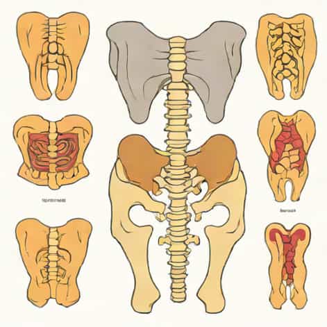
The Young and Burgess classification divides pelvic fractures into three primary types based on the mechanism of injury lateral compression (LC), anteroposterior compression (APC), and vertical shear (VS). Each type is further subdivided to describe the severity and specific characteristics of the fracture. This system allows clinicians to quickly assess the potential for pelvic instability and associated injuries, which is important because pelvic fractures often involve significant blood loss and may threaten life. Unlike older classification systems that focused solely on anatomical description, Young and Burgess emphasizes the forces responsible for the injury, providing both diagnostic and therapeutic value.
Key Principles
- Focuses on the mechanism of injury lateral, anteroposterior, or vertical force.
- Links fracture pattern to pelvic stability and risk of hemorrhage.
- Subdivides fractures into grades based on severity and displacement.
- Guides treatment decisions, including surgical intervention versus conservative management.
- Improves communication among trauma and orthopedic teams regarding fracture severity.
Lateral Compression (LC) Fractures
Lateral compression fractures occur when force is applied from the side of the pelvis, often during side-impact motor vehicle collisions. The force causes internal rotation of the hemipelvis, leading to characteristic fracture patterns. LC fractures are generally subdivided into three types LC-I, LC-II, and LC-III, each reflecting increasing severity and involvement of the pelvic ring and posterior structures. These fractures are often associated with internal organ injuries and can cause significant morbidity if not managed appropriately. Most LC-I and LC-II fractures are considered stable or partially stable, while LC-III fractures often result in rotational and vertical instability.
Subtypes of Lateral Compression
- LC-I Sacral compression on the same side as the lateral impact; stable posterior ring fracture.
- LC-II LC-I fracture plus posterior ilium fracture with rotational instability.
- LC-III Windswept pelvis with contralateral open-book pattern, leading to rotational and vertical instability.
Anteroposterior Compression (APC) Fractures
Anteroposterior compression fractures, commonly called open-book fractures, result from force applied to the front of the pelvis, such as head-on vehicle collisions. These injuries cause external rotation of the hemipelvis and can lead to diastasis of the pubic symphysis. APC fractures are classified into three types APC-I, APC-II, and APC-III. The severity increases from mild widening of the pubic symphysis to complete disruption of the anterior and posterior pelvic ring. APC fractures carry a high risk of hemorrhage due to disruption of pelvic vessels and often require prompt intervention to stabilize the pelvis.
Subtypes of Anteroposterior Compression
- APC-I Mild symphyseal diastasis (<2.5 cm); generally stable.
- APC-II Significant symphyseal widening (>2.5 cm) with anterior sacroiliac ligament disruption; rotationally unstable.
- APC-III Complete anterior and posterior pelvic ring disruption; both rotationally and vertically unstable, high risk of hemorrhage.
Vertical Shear (VS) Fractures
Vertical shear fractures occur when a vertical force, such as a fall from a significant height, acts on one hemipelvis. The force causes upward displacement of the hemipelvis, disrupting both the anterior and posterior pelvic ring. These fractures are highly unstable and are associated with severe hemorrhage and soft tissue injury. Vertical shear fractures typically require surgical stabilization due to their instability and the risk of life-threatening complications. Understanding the mechanism of injury helps clinicians anticipate potential vascular and organ injuries that may accompany VS fractures.
Characteristics of Vertical Shear Fractures
- Vertical displacement of one hemipelvis.
- Complete disruption of anterior and posterior pelvic structures.
- High risk of vascular injury and hemorrhage.
- Often associated with soft tissue and organ trauma.
- Requires surgical fixation for stability and functional recovery.
Clinical Relevance of the Classification
The Young and Burgess classification is clinically relevant because it helps guide immediate and long-term management of pelvic fractures. By identifying the type of fracture and its stability, trauma teams can determine the urgency of pelvic stabilization, the need for blood transfusions, and whether surgical intervention is required. It also assists in predicting associated injuries, such as bladder, urethral, or vascular trauma, which are common with severe pelvic fractures. In addition, the classification helps in documenting injury severity for research, communication among healthcare providers, and assessing prognosis.
Benefits in Clinical Practice
- Guides emergency stabilization and treatment decisions.
- Predicts risk of hemorrhage and associated injuries.
- Helps prioritize surgical versus non-surgical management.
- Facilitates clear communication between orthopedic surgeons and trauma teams.
- Supports research and outcome tracking for pelvic trauma.
Imaging and Diagnosis
Accurate classification of pelvic fractures using the Young and Burgess system relies on high-quality imaging. X-rays provide initial assessment, while computed tomography (CT) scans offer detailed visualization of the pelvic ring and posterior structures. CT imaging is particularly useful in identifying subtle fractures, posterior sacroiliac disruptions, and vertical displacement. MRI may also be used in specific cases to assess soft tissue and ligamentous injury. Combining clinical examination with imaging findings ensures accurate classification, which is critical for effective management and reducing complications.
Imaging Techniques
- Plain radiographs (X-rays) Anteroposterior, inlet, and outlet views.
- CT scans Detailed assessment of posterior pelvic ring and displacement.
- MRI Evaluation of ligaments and soft tissue in complex cases.
- Fluoroscopy Sometimes used intraoperatively for real-time assessment.
- Physical examination Complements imaging for stability evaluation.
The Young and Burgess classification of pelvic fractures provides a comprehensive framework for understanding and managing complex pelvic injuries. By focusing on the mechanism of injury, fracture pattern, and pelvic stability, the system allows clinicians to predict complications, guide treatment decisions, and improve patient outcomes. Lateral compression, anteroposterior compression, and vertical shear fractures each present unique challenges, with varying degrees of instability and associated risks. Accurate classification, combined with proper imaging and clinical assessment, ensures that patients with pelvic fractures receive timely, appropriate, and effective care, ultimately improving both survival and functional recovery.