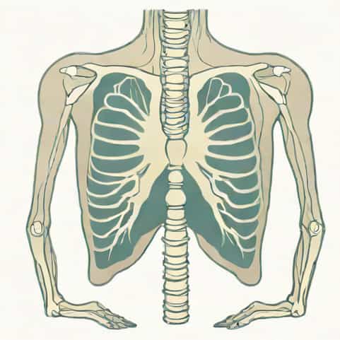
Optic neuritis is a condition that involves inflammation of the optic nerve, which can lead to visual disturbances, pain, and in some cases, long-term vision loss. While optic neuritis primarily affects the eyes and the nervous system, chest X-rays may sometimes be ordered as part of a broader diagnostic approach, particularly when underlying systemic conditions are suspected. Understanding the connection between optic neuritis and chest imaging can be important in evaluating patients for autoimmune disorders, infections, or inflammatory conditions that may involve multiple organ systems. Exploring this topic provides insight into why imaging tests such as chest X-rays are sometimes included in the workup of neurological symptoms and how healthcare providers use these tools to form a comprehensive assessment.
What Is Optic Neuritis?
Optic neuritis occurs when the optic nerve, which transmits visual information from the eye to the brain, becomes inflamed. This inflammation can result in sudden or progressive vision loss, typically in one eye, along with pain during eye movement. The condition may be associated with autoimmune disorders like multiple sclerosis (MS), infections, or other inflammatory diseases. Optic neuritis can affect people of all ages, though it is most commonly observed in young adults and is more prevalent in women.
Symptoms of Optic Neuritis
- Sudden vision loss in one eye or both eyes in rare cases
- Pain, especially when moving the affected eye
- Blurred vision or reduced color perception
- Flashing lights or flickering in vision
- Temporary vision deterioration over days to weeks
Because optic neuritis can be an early sign of conditions like multiple sclerosis, a thorough diagnostic evaluation is crucial. This often includes neurological exams, blood tests, MRI of the brain and optic nerve, and sometimes additional imaging such as chest X-rays to rule out other causes.
Role of Chest X-Ray in Optic Neuritis Diagnosis
While optic neuritis itself affects the optic nerve, a chest X-ray can provide information about underlying systemic conditions that may be contributing to the inflammation. Chest X-rays are particularly useful in identifying infections, inflammatory conditions, or tumors that could indirectly affect the optic nerve. For example, sarcoidosis, a condition characterized by the growth of tiny clusters of inflammatory cells in various organs, often shows abnormalities in the lungs visible on a chest X-ray. Detecting such issues can guide further evaluation and treatment of optic neuritis.
Conditions Identified on Chest X-Ray Relevant to Optic Neuritis
- SarcoidosisGranulomas in the lungs can indicate systemic inflammation that may affect the optic nerve.
- Tuberculosis or other infectionsInfections in the lungs may trigger immune responses that lead to optic nerve inflammation.
- Lymphoma or tumorsCertain cancers may present with both ocular and thoracic manifestations.
- Autoimmune conditionsSome autoimmune diseases affecting the lungs can also involve neurological symptoms.
Chest X-rays, therefore, are not used to diagnose optic neuritis directly, but they provide a valuable perspective on potential systemic causes. A normal chest X-ray does not rule out optic neuritis but can help narrow down the list of possible underlying conditions.
Diagnostic Process for Optic Neuritis
The evaluation of optic neuritis typically involves multiple steps. Medical history and physical examination are the first steps, focusing on visual symptoms, eye movement pain, and any neurological complaints. An ophthalmologic examination may include visual acuity tests, color vision tests, and fundoscopy to assess the optic nerve head. MRI is the most informative imaging modality for optic neuritis, showing inflammation or demyelination of the optic nerve.
Additional Tests in Diagnosis
- Blood testsTo check for infections, autoimmune markers, or vitamin deficiencies.
- Chest X-rayTo identify lung involvement, sarcoidosis, or other systemic causes.
- Cerebrospinal fluid analysisTo assess for multiple sclerosis or other inflammatory conditions.
- Visual evoked potentialsTo measure electrical activity in the optic nerve in response to visual stimuli.
Integrating chest X-ray results with other tests helps clinicians determine whether optic neuritis is isolated or part of a broader systemic issue. For example, bilateral hilar lymphadenopathy on a chest X-ray may suggest sarcoidosis as the underlying cause.
Treatment and Management
Once diagnosed, optic neuritis is typically managed with corticosteroids to reduce inflammation and accelerate recovery of vision. The treatment plan may also include addressing the underlying systemic condition, such as prescribing medications for autoimmune diseases or infections identified via chest X-ray or other tests. Recovery varies; many patients regain significant vision over weeks to months, although some may experience residual visual deficits.
Monitoring and Follow-Up
- Regular ophthalmologic assessments to track vision recovery
- MRI follow-up to monitor optic nerve health
- Systemic evaluations if chest X-ray abnormalities indicate conditions like sarcoidosis or tuberculosis
- Neurological monitoring for potential progression to multiple sclerosis
Effective follow-up ensures that both optic neuritis and any associated systemic condition are managed appropriately. Coordination between ophthalmologists, neurologists, and pulmonologists may be necessary, particularly when chest X-ray reveals lung involvement.
Optic neuritis is an inflammatory condition affecting the optic nerve that can lead to vision loss and eye pain. While chest X-rays do not directly diagnose optic neuritis, they play a valuable role in identifying systemic conditions such as sarcoidosis, infections, or tumors that may be contributing factors. Combining chest X-ray findings with MRI, blood tests, and clinical evaluations allows healthcare providers to form a comprehensive diagnosis and treatment plan. Understanding the relationship between optic neuritis and chest X-ray results helps ensure that both ocular and systemic conditions are addressed, improving patient outcomes and guiding effective management of inflammation and associated health risks.