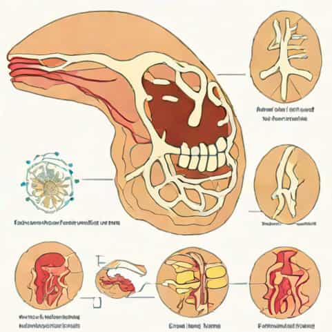
Bone formation is a fascinating biological process that allows the human body to develop, grow, and repair itself. One of the main methods by which bones form is called endochondral ossification. This process involves the gradual transformation of cartilage into bone and is essential for the development of most of the skeleton, especially long bones such as the femur, tibia, and humerus. Understanding the steps of endochondral ossification helps students, healthcare professionals, and anatomy enthusiasts appreciate how the body constructs its strong yet dynamic framework from early development through adulthood.
Understanding Endochondral Ossification
Endochondral ossification begins during fetal development and continues as a person grows. Unlike intramembranous ossification, which directly converts mesenchymal tissue into bone (as seen in flat bones like the skull), endochondral ossification first forms a cartilage model. This cartilage serves as a template that is gradually replaced by bone tissue. The process is highly organized and involves a sequence of cellular and structural changes that ensure bones develop with the correct shape, length, and strength.
The Major Steps of Endochondral Ossification
To better understand how cartilage turns into bone, it’s important to follow the process step by step. Each stage builds upon the previous one, involving specific cells and biochemical signals that coordinate the transformation. The steps of endochondral ossification can be summarized as follows
- Formation of a cartilage model
- Development of the bone collar
- Formation of the primary ossification center
- Appearance of the medullary cavity
- Development of secondary ossification centers
- Formation of articular cartilage and epiphyseal plate
Step 1 Formation of the Cartilage Model
The first step in endochondral ossification begins with the development of a cartilage model. Mesenchymal cells, which are unspecialized stem cells, group together in the shape of the future bone. These cells then differentiate into chondroblasts, specialized cells responsible for producing cartilage. The chondroblasts secrete a flexible matrix made of collagen and proteoglycans, forming a hyaline cartilage model that mimics the shape of the final bone.
As the cartilage model grows, chondroblasts become trapped within the matrix and mature into chondrocytes. The cartilage expands in length and width through two types of growth interstitial growth (from within) and appositional growth (on the outer surface). At this stage, the structure is still entirely made of cartilage, not bone, but it establishes the blueprint for the skeleton’s later ossification.
Step 2 Development of the Bone Collar
Once the cartilage model has reached a certain size, changes begin in the outer layer. The perichondrium, a membrane surrounding the cartilage, transforms into a periosteum a bone-producing layer. Cells in this region differentiate into osteoblasts, which are bone-forming cells. These osteoblasts start producing a thin layer of compact bone around the midsection of the cartilage model, known as the bone collar.
This bone collar provides strength and structure to the developing bone, but it also limits the diffusion of nutrients to the chondrocytes inside the cartilage. As a result, the chondrocytes in the center begin to enlarge and eventually die, triggering the next stage of the process. The bone collar is an essential early step because it signals the transition from a cartilage model to true bone tissue.
Step 3 Formation of the Primary Ossification Center
As the chondrocytes in the center of the cartilage model die, the surrounding matrix begins to break down, creating small cavities. Meanwhile, blood vessels from the periosteum invade the center of the cartilage, carrying with them osteoblasts and osteoclasts. This invasion marks the formation of the primary ossification center, which usually appears in the diaphysis (the shaft of the long bone).
Osteoblasts begin to deposit bone matrix in the empty spaces left by the dying cartilage. This process replaces the cartilage with spongy bone tissue. The bone continues to grow toward both ends of the cartilage model, as osteoblasts keep depositing new matrix while osteoclasts remove old material to shape the marrow cavity. This stage is crucial because it establishes the first true bone tissue within the developing structure.
Step 4 Formation of the Medullary Cavity
As ossification progresses within the diaphysis, the central spongy bone tissue is broken down by osteoclasts, forming the medullary (marrow) cavity. This hollow space will later house bone marrow, which is responsible for producing blood cells. The bone collar surrounding the shaft thickens, giving the bone additional support.
This stage not only creates space for the bone marrow but also marks the beginning of bone remodeling a lifelong process where old bone is replaced by new bone tissue. During this phase, blood vessels continue to expand through the cavity, ensuring the developing bone receives the nutrients and oxygen it needs for further growth.
Step 5 Development of Secondary Ossification Centers
While the primary ossification center continues to develop in the diaphysis, secondary ossification centers begin to form in the epiphyses (the ends of the bone). This typically occurs shortly before or after birth. Similar to the earlier stage, blood vessels invade the epiphyses, bringing osteoblasts and osteoclasts that start replacing cartilage with bone tissue.
Unlike the diaphysis, the secondary ossification centers do not form a medullary cavity. Instead, they retain some cartilage on the surface, which will later become the articular cartilage. This cartilage allows smooth movement between bones in a joint, reducing friction and preventing wear. The formation of secondary ossification centers completes the basic structure of the bone, although growth continues through childhood and adolescence.
Step 6 Formation of Articular Cartilage and the Epiphyseal Plate
The final stage of endochondral ossification involves the development of the articular cartilage and the epiphyseal (growth) plate. The thin layer of cartilage covering the epiphyses remains as articular cartilage, which helps in joint movement. Between the diaphysis and each epiphysis lies the epiphyseal plate, a region of hyaline cartilage responsible for longitudinal bone growth.
As a child grows, new cartilage is continuously produced at the epiphyseal plate, while older cartilage is replaced by bone. This process causes the bone to lengthen. Once growth stops typically in early adulthood the epiphyseal plate is entirely replaced by bone, leaving behind a visible line called the epiphyseal line. At this point, the bone has reached its full adult length and the process of endochondral ossification is complete.
Summary of the Steps in Order
To summarize the sequence clearly, here is the order of the steps of endochondral ossification
- Mesenchymal cells form a cartilage model.
- The perichondrium transforms into periosteum and forms a bone collar.
- Chondrocytes in the center die, and a primary ossification center forms in the diaphysis.
- The medullary cavity develops as bone remodeling begins.
- Secondary ossification centers appear in the epiphyses.
- Articular cartilage and the epiphyseal plate remain for joint function and continued growth.
Each step is tightly regulated by genetic and hormonal signals to ensure that bones develop properly. Disturbances in any of these steps can lead to skeletal deformities or growth disorders.
The Importance of Endochondral Ossification
Endochondral ossification is not only essential for the development of long bones but also for bone repair. When a bone fractures, the body partially repeats this process. A cartilage callus forms at the break site, which is then replaced by bone through ossification. This highlights how the mechanism remains active throughout life, not just during early development.
Additionally, understanding this process provides valuable insight into various medical conditions such as dwarfism, rickets, and osteogenesis imperfecta. Each of these disorders can disrupt normal bone growth or mineralization, emphasizing the importance of proper endochondral development for skeletal health.
Endochondral ossification is a beautifully coordinated process that transforms cartilage into bone through a series of well-defined steps. From the initial formation of a cartilage model to the creation of secondary ossification centers and the closing of the growth plates, each phase plays a vital role in building strong, functional bones. This process underlies the growth of most bones in the human body and continues to influence repair and remodeling throughout life. Understanding the order of these steps not only deepens our appreciation for human biology but also provides essential knowledge for medical, biological, and anatomical studies.